Friday July 13, 2007
Dian took us into the electron microscope room to use the scanning electron microscope and to see the transmission electron microscope. Dian had prepared her nanotubes the day before. The nanotubes self assemble when the temperatures they are exposed to vary (this sounds easy and rather sloppy, but like everything that is done here, it is neither!). She placed her sample on a metal sample holder (looks like a circular, thick brass plate), adhering it with double sided carbon tape , chosen because it can conduct. We take the mounted sample into another room, where the sample is spatter coated with a layer of gold between 1 and 2 nanometers thick.
The sample is then brought back to the Electron Microscopy Lab and it is loaded onto the SEM sample insertion device.
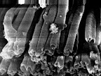
The picture immediately above on the right shows the sample inside the SEM sample chamber. At the time of this photo, Dian has evacuated the SEM sample chamber, and the beam has been centered. Control knobs are used to move the sample horizontally and vertically within the chamber. The contrast and magnification knobs are used to obtain the sharpest possible image at the desired magnification. We located tubes at a 40,000 magnification. The image below left is a low magnification of the tube sample, and to the right is a high magnification showing tubes.
All of us had an opportunity to move the sample, adjust the magnification, and take pictures. These pictures are digital, so they are easily transferable as files. The TEM is quite old however, and all of the pictures are taken on photographic film and must be chemically developed (old school indeed). Using the SEM, Dian is trying to see how smooth the surfaces of the tubes are.

Wednesday, July 18, 2007
Scanning Electron Microscope, Dr. Hashimoto
Posted by
Chaug Biology Research
at
11:49 AM
![]()
Subscribe to:
Post Comments (Atom)


No comments:
Post a Comment